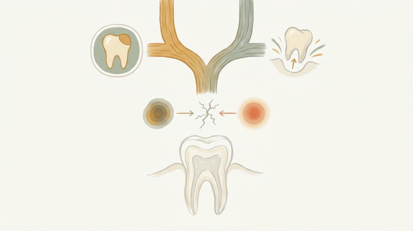
A dentist chooses a filling when enough healthy tooth structure remains to support a restoration and the decay has not destroyed the tooth's foundation. Extraction becomes the answer when decay or a fracture extends too far below the gumline, too little sound structure is left to hold a repair, or a crack runs down the root. The decision is made by combining a hands-on exam, your history, and targeted X-rays — not by looking at symptoms alone.
If you are in Vineland and you have been putting off a sore tooth because you are afraid the answer will be "pull it," the honest news is that most teeth we see are savable. The teeth that aren't usually tell us so on the X-ray before the patient ever feels the full picture. Here is how the call actually gets made, step by step.
You usually can't know on your own — the deciding factors live below the surface. A dentist measures how much healthy tooth is left, whether decay or a crack has reached the pulp or the root, and whether the bone and gum still support the tooth. A filling fits small-to-moderate decay; extraction is reserved for teeth that can no longer hold a restoration or whose root is split.
Symptoms are a starting point, not a verdict. A tooth can ache sharply and still be fillable, and a tooth with mild symptoms can turn out to be non-restorable once imaged. That is why guesswork at home almost always underestimates one direction or the other.
Dentists weigh four things: the amount of remaining healthy tooth structure, the depth and spread of the decay or fracture, whether the pulp (the nerve chamber inside the tooth) is involved, and the health of the bone and gum around the tooth. Each factor can push the decision toward a filling, a root canal plus restoration, or an extraction — in that order of preference.
A filling repairs cavities and minor chips or cracks by removing the decayed portion of the tooth before it gets too close to the pulp and replacing it with a specially formulated material, which stops the decay and gives the tooth a chance to heal. That is the whole logic of a filling in one sentence. The moment any of those four conditions fails, the plan has to change.
At Vineland Dental, our team walks through those four checkpoints with you on the screen so the recommendation isn't a mystery — the practice's standard is that no patient leaves without understanding exactly what is happening and why.
A cavity is too big for a filling when the decay has eaten through so much of the tooth that a filling would have nothing solid to bond to, or when the decay has tunneled into the pulp and then down into the root or jawbone. At that point the choice shifts to a root canal with a crown (if structure remains) or extraction (if it doesn't).
Here is the progression dentists are tracking. Cavities localized to the hard outer enamel can take years to progress, or may not progress at all, which is why not every spot on an X-ray gets drilled on day one. "Incipient cavities," or surface-level demineralization of the enamel, have not yet penetrated to the second, much softer layer of the tooth — and the NIDCR notes the tooth decay process happens over time and can be interrupted or even reversed to avoid a cavity. Early mineral loss that hasn't formed a hole can sometimes be managed without drilling, for example with fluoride and preventive care.
Once decay crosses into that softer inner layer, it moves faster. When bacteria-produced acid finally creates a tunnel to the center of the tooth housing the nerve, bacteria gain straight-line access down the nerve to the root and ultimately into the jaw bone. That is the threshold at which a simple filling is no longer the honest answer.
No. Decay that reaches the pulp does not automatically mean extraction — root-canal treatment can remove infected or irreversibly damaged pulp and seal the tooth, which can then be restored if enough sound structure remains. Extraction becomes the recommendation only when decay or a break extends too far below the gumline, too little sound structure remains to retain a restoration, or a crack runs down the root.
Advanced gum disease that has left a tooth poorly supported or very loose can also make a restoration not worthwhile, which is why dentists assess the tooth and its surrounding tissues rather than looking only at the visible cavity. A perfectly fillable crown of a tooth is still a lost cause if the bone underneath has melted away.
Because the deciding details — how deep the decay runs, whether it has reached the pulp, whether a crack has reached the root, and whether the bone is still intact — are not visible to the naked eye. ADA imaging guidance identifies bitewing X-rays as useful for suspected decay between back teeth that cannot be seen clinically, and periapical views for certain hidden decay or root concerns.
Current ADA guidance (January 2026) emphasizes examining the patient and reviewing relevant history and prior images before ordering X-rays; imaging should be based on clinical need, not taken on a fixed schedule. In practice that means your dentist should look in your mouth and ask about your history first, then order the specific views that will actually change the plan — not a blanket set.
If the tooth is restorable, the two broad categories are direct fillings (shaped in the mouth in one visit) and indirect fillings (made in a lab or mill and bonded in a second visit). Your dentist will let you know how many visits your specific restoration requires.
Materials matter for how the repair looks and how long it lasts. Depending on the material used, some dental fillings can last up to 20 years.
In Vineland, the practical wrinkle is access. Getting evaluated early, especially before the holiday stretch when schedules tighten, is often what keeps a tooth in the filling column.
Vineland Dental sits downtown near the Landis Avenue business district, and the team includes Spanish-speaking staff members Tania and Ashley, which matters in a city where a large share of households speak Spanish at home. Office Manager Sandy Stites has been part of the practice for 26 years, going back to when it was known as Vineland Dental Arts, and the practice itself has been part of the Vineland community for over 26 years. Lead dentist Dr. Walter Hinman graduated from Temple University's dental school in 2017 and served as a US Army dentist from 2017 to 2021, including a nearly six-month deployment aboard the USNS Comfort in 2019.
No — the factors that decide it live below the enamel, where only X-rays can see them. Pain level isn't a reliable guide: a sharp ache can come from a fillable cavity, and a nearly painless tooth can turn out to be split at the root. A dental exam plus targeted bitewing or periapical X-rays is the only way to know which category your tooth falls into.
Not necessarily. Cavities confined to the hard outer enamel can take years to progress, or may not progress at all, which is why every spot isn't drilled on day one. Early mineral loss that hasn't yet formed a hole can sometimes be managed without drilling, for example with fluoride and preventive care. Once decay crosses into the softer inner layer of the tooth, it moves faster and a filling becomes time-sensitive.
No. Decay reaching the pulp does not automatically mean extraction — a root canal can remove the infected or irreversibly damaged pulp and seal the tooth, which can then be restored with a crown if enough sound structure remains. Extraction is reserved for teeth where decay or a break extends too far below the gumline, too little structure remains to hold a restoration, or a crack runs down the root.
Your dentist will tell you which type of filling suits the size and location of your cavity and how many visits it will require.
It depends on the material and on how the tooth is used, but some dental fillings can last up to 20 years. Different filling materials each have their own strengths depending on the tooth's location and bite load. Regular checkups catch wear before a filling fails.
Because the decisive details aren't visible by eye. ADA imaging guidance identifies bitewing X-rays as useful for suspected decay between back teeth that cannot be seen clinically, and periapical views for certain hidden decay or root concerns. Current ADA guidance (January 2026) emphasizes examining the patient and reviewing relevant history and prior images before ordering X-rays, with imaging based on clinical need rather than a fixed schedule.
Tell the front desk when you book. A good practice will slow down, show you what the X-ray is showing, and talk through the options before anything is decided. Vineland Dental's stated standard is that no patient leaves without understanding exactly what is happening and why, and the team includes Spanish-speaking staff members Tania and Ashley for patients who are more comfortable in Spanish.
Fillings win whenever the tooth still has enough honest structure to hold one and the pulp and root are intact. Root canals rescue teeth whose pulp is damaged but whose foundation is sound. Extraction is the right answer when the foundation itself is gone — decay below the gumline, a crack down the root, or bone loss that no restoration can fix. The only reliable way to know which bucket your tooth is in is an exam with the right X-rays.
Readers in Vineland, NJ who want a straight answer on a specific tooth can reach Vineland Dental at (856) 205-9500 or https://vinelanddental.com to schedule an evaluation. Bringing the tooth in sooner rather than later is usually what keeps the decision in the filling column.